A black spot on a tooth is one of those discoveries that stops people cold during their morning brushing routine, and the first question is almost always the same: Is it a cavity? At Dillon Family Dentistry on East Lancaster Avenue in Bryn Mawr, PA, Dr. David Dillon sees this concern regularly from Main Line patients, and the answer is never one-size-fits-all. A black spot can be a surface stain that needs cleaning, an early cavity that needs a small filling, or, in some cases, a sign of more advanced decay requiring prompt attention. The only way to know which it is, and to get it treated before it becomes a bigger problem, is to have it evaluated by your dentist.
You are brushing your teeth, you catch a glimpse in the mirror, and there it is: a small black or dark spot on one of your teeth. Maybe it is on a back molar where you could not see it easily before, or maybe it is somewhere more visible that has you genuinely concerned. Your first question is the right one: Is it a cavity?
I am Dr. David Dillon, and from our office on East Lancaster Avenue here in Bryn Mawr, I can tell you that this is one of the most common questions I get from patients across the Main Line. The honest answer is: it might be a cavity, and it might not be. A black spot has several possible causes, ranging from surface staining that requires nothing more than a professional cleaning to early tooth decay that can be treated with a small filling if caught now, to more advanced decay that needs more involved care. What it is not, ever, is something to ignore or put off having evaluated. Here is exactly what you need to know.
What Is a Black Spot on a Tooth? The Four Main Possibilities
Not every dark mark on a tooth represents the same problem, and the distinction matters enormously for how it should be treated. Here are the four most common causes I see at my Bryn Mawr practice:
1. Tooth Decay (Cavity)
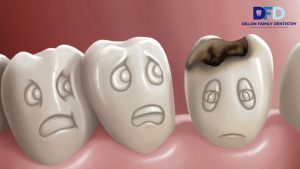
Dental caries, commonly called a cavity, is the most serious possibility and the one patients are most concerned about, rightly so. A cavity forms when bacteria in the mouth produce acid that demineralizes and destroys tooth enamel. In the early stages, this produces a white or chalky spot. As decay progresses through the enamel and into the dentin below, the affected area darkens to brown and then black as the destroyed tooth structure stains.
The critical thing to understand is that early-stage cavities, even ones that appear as visible black spots, are often completely painless. Tooth pain from a cavity typically does not develop until decay reaches the inner pulp, where the nerve lives. By that stage, a simple filling is no longer sufficient, and root canal therapy is likely needed. A small black spot that is an early cavity is a routine filling. The same spot ignored for another year may be a crown or a root canal. That is not an exaggeration. That is the documented progression of untreated decay.
2. Surface Staining (Extrinsic Discoloration)
Not every black mark is decay. Heavily pigmented foods and beverages, including coffee, black tea, red wine, and dark sauces, deposit chromogens on the enamel surface over time. These deposits can concentrate in specific areas, particularly in the natural grooves and pits of the chewing surfaces of molars, producing what looks exactly like a small dark cavity to the naked eye. Tobacco use, both smoked and chewed, is among the most aggressive causes of this type of dark surface staining.
The good news about extrinsic staining is that it is purely cosmetic and has no clinical significance for tooth health. A professional cleaning removes most surface staining, and teeth whitening addresses the rest. The challenge is that staining and early decay look nearly identical without a clinical probe and X-ray. This is why a professional evaluation is always the right move when you notice a new dark spot, even if you suspect it is just a stain.
3. Black Tartar at or Near the Gum Line
You may notice a dark line or series of dark spots, specifically at the gum line on the inside surfaces of the lower front teeth or near the gum margins on other teeth. This is almost always black tartar, also called subgingival calculus. Regular yellowish tartar is calcified plaque. Black tartar forms when plaque calcifies below or at the gum line and becomes stained by blood pigments from the inflamed gum tissue and bacterial metabolic products. It is associated with gum disease and is significantly more difficult to remove than regular tartar.
Black tartar can only be removed by professional cleaning, specifically the type of deep cleaning we perform at our Main Line family dental practice. It cannot be brushed away at home. Its presence is also a signal that we need to assess the health of the gum tissue in that area.
4. Staining from Old Dental Work
If you have had fillings or other restorations, particularly older amalgam (silver) fillings, the metal can leach into the surrounding tooth structure over time and create a gray or dark discoloration in the tooth around or beneath the restoration. This is called amalgam tattooing of the tooth and is cosmetic in nature, not a sign of active decay. However, any old restoration warrants evaluation at a dental visit, as older fillings can fail at the margins and allow new decay to develop beneath or around them.
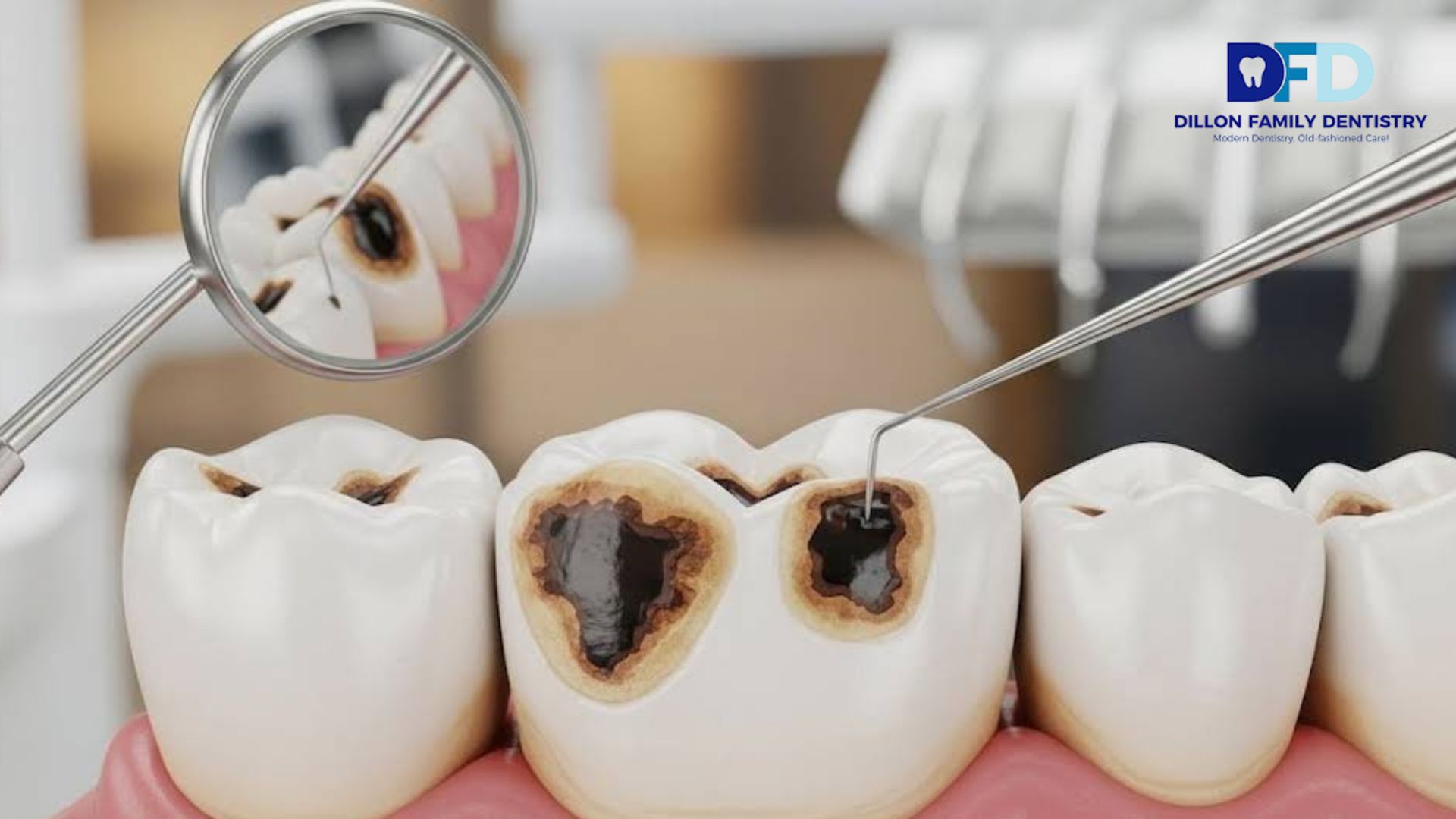
How to Tell a Stain from a Cavity: What I Look for in the Exam Room
This is the question I hear most often, and I want to be direct: you cannot reliably tell the difference between a surface stain and an early cavity based on visual inspection alone, even with a mirror and good lighting. Both produce black or dark brown spots. Both may be painless. The spot location gives some clues, but it is not definitive.
Here is what we do at Dillon Family Dentistry to make the distinction:
- Clinical probing: I gently explore the spot with a dental explorer. Healthy enamel and surface staining feel smooth and hard. Decay has a characteristic texture: slightly soft or sticky, and the explorer may get caught in a pit. This tactile assessment is one of the first and most informative diagnostic steps.
- Digital X-rays: Bitewing and periapical X-rays reveal decay between teeth and beneath existing restorations that cannot be seen at all during visual inspection. We use digital radiography at our Bryn Mawr office, which reduces radiation exposure compared to traditional film X-rays and provides images I can review with you on-screen immediately.
- Visual examination with magnification: Using a dental loupe or microscope, we can assess the texture, margins, and surface characteristics of a suspicious spot in detail, which is impossible with the naked eye.
- Transillumination: In some cases, shining a focused light through the tooth reveals internal decay patterns that are not visible otherwise.
With these two tools, I can come to an answer that is not open to question. Stained wood has some degree of “slickness” when felt underhand. Wood with ongoing decay or other structural involvement has a characteristic consistency of “softness”, or as I call it, a “toilet tissue” feel. If there is any uncertainty about the appearance of a potential problem, where there is a “spot” that has an unusual or questionable appearance, I will evaluate the area again at the next appointment for an injection rather than “drilling” on it unnecessarily. As a rule of thumb, the most conservative procedure is taken for any tooth, such that the health of the tooth is maintained at all times.
The Stages of Tooth Decay: Why Timing Changes Everything
Tooth decay does not arrive fully formed. It progresses through distinct stages, each of which has implications for how it must be treated. Understanding the stages helps explain why I am so emphatic about early detection.
Stage 1: Demineralization (White Spot Lesion)
Early signs of enamel decay occur before a tooth develops into a cavity. These early decay signs can be seen as a white or chalky spot. If treated early with fluoride and good hygiene, some enamel loss due to acid attack can occasionally be stopped without drilling. During the earliest stage of decay, the enamel is still intact and capable of being restored with preventive measures.
Stage 2: Enamel Decay
Once the acid has fully penetrated the enamel surface, a true cavity exists. The area becomes brown or black and slightly soft. Treatment is a composite filling, which we place in the same appointment as the diagnosis in most cases. These are small, conservative restorations that preserve as much natural tooth structure as possible. We have placed only metal-free, tooth-colored composite fillings at our Bryn Mawr office for over 25 years because they bond to the tooth structure and support what remains, rather than creating the stress that older amalgam restorations can impose.
Stage 3: Dentin Decay
Enamel is the hardest substance in the body. Dentin, the layer beneath it, is softer and more porous, and once decay reaches it, progression accelerates significantly because acid moves through dentin much faster. At this stage, sensitivity, particularly to cold and sweets, often begins. Treatment is still a filling, but a larger one. Depending on how much tooth structure has been lost, a dental crown may be needed to restore proper form and function.
Stage 4: Pulp Involvement
When decay reaches the pulp chamber, the inner space containing the tooth’s nerve and blood supply, the character of the pain changes from sensitivity to persistent, throbbing pain. At this stage, root canal therapy is required to remove the infected or inflamed pulp tissue before the tooth can be restored with a crown. This is significantly more involved and more expensive than a filling. It is the stage that patients reach when they ignore a painless black spot for too long.
Stage 5: Abscess and Tooth Loss
Untreated pulp infection leads to a dental abscess, a pocket of bacterial infection that can spread to the surrounding bone and tissue. At this point, extraction may be the only viable option. This is the outcome that begins as a small black spot noticed during morning brushing.
Location Matters: What a Black Spot’s Position Tells Us
Where on the tooth you find a dark spot provides diagnostic clues, even before we perform the formal exam:
On the Chewing Surface (Occlusal)
The deep grooves and pits of the biting surfaces of molars are the most common site for cavities in adults and children alike. These grooves are narrow enough that toothbrush bristles cannot fully clean them, and food particles and bacteria accumulate. A black spot or pit in the center of a molar’s chewing surface is highly suspicious for decay, though it can also be staining concentrated in those grooves. This is the site where dental sealants provide the most protective benefit.
Between Teeth (Interproximal)
Cavities between teeth are often invisible to visual inspection entirely and show up only on X-rays as dark shadows between the tooth roots. If you notice a dark shadow at the contact point between teeth, or if you have pain when flossing, an interproximal cavity may be the cause. This is another reason that regular X-rays are not optional. They catch decay in locations that are simply not visible any other way.
Near the Gum Line
Dark spots at or just below the gum line are more commonly black tartar than decay in patients with intact root surfaces, but in patients who have experienced gum recession, root surface decay is a real concern. Root surfaces do not have a protective enamel layer, making them significantly more vulnerable to decay. A dark spot on the exposed root surface near the gum line needs careful evaluation to distinguish tartar from root caries.
On the Smooth Surfaces
Black spots on the smooth buccal or labial surfaces of teeth, the cheek-facing or lip-facing surfaces, are more likely to be staining or tartar than cavities. Smooth-surface caries are less common and tend to develop in areas of plaque accumulation near the gum margin.
Cavity Treatment Options in Bryn Mawr, PA
Once we have established that a black spot represents decay rather than staining, here is what treatment looks like at Dillon Family Dentistry:
Composite Fillings
For early to moderate cavities confined to the enamel and upper dentin layers, a tooth-colored composite resin filling is the standard of care. The decayed portion is removed, the cavity is cleaned, and the composite material is bonded to the tooth in layers and hardened with a curing light. We match the composite shade to the surrounding tooth precisely so the restoration is virtually invisible. We have been placing metal-free composite fillings exclusively for over 25 years at our Bryn Mawr office. Learn more about our family dental services on our family dental page.
Dental Crowns
When a cavity is extensive enough that the remaining tooth structure cannot support a filling reliably, or when a cusp has fractured, a dental crown is the appropriate restoration. The crown covers the entire visible tooth, distributing biting forces evenly and protecting the underlying structure. We place beautiful, metal-free ceramic crowns at our office. See our dental implants and crowns page for more information.
Root Canal Therapy
When decay has reached the pulp, root canal therapy is required before the tooth can be restored. I want to address the reputation directly: modern root canal treatment under proper local anesthesia is no more uncomfortable than a filling appointment. We remove the infected pulp tissue, disinfect and shape the canals, seal them, and restore the tooth with a crown. Most of our root canals are completed in a single visit.
Fluoride Remineralization for Early Lesions
For white spot lesions and very early enamel demineralization that has not yet progressed to a true cavity, professional fluoride treatment, combined with improved home fluoride use and dietary modifications, can sometimes arrest and partially reverse the lesion without any drilling. This approach is only appropriate for the earliest stage of decay and requires close monitoring. It is one of the reasons we encourage patients to come in regularly rather than waiting until they have symptoms.
Preventing Cavities and Black Spots on Teeth
Prevention is dramatically simpler and less expensive than treatment, and the strategy is well established:
- Brush with fluoride toothpaste for two minutes twice daily, paying particular attention to the gum line and the pits and grooves of back teeth.
- Floss daily. Interproximal cavities develop in the spaces between teeth that no toothbrush reaches. Flossing is the only home care measure that prevents decay in those spaces.
- Reduce sugar and refined carbohydrate consumption, especially between meals. The acid produced by bacteria feeding on sugar attacks enamel within minutes of consumption and continues for 20 to 30 minutes afterward.
- Drink water with fluoride. The public water supply in most of the Main Line area, including Bryn Mawr, Haverford, and Ardmore, contains fluoride that provides measurable protection against tooth decay at the community level.
- Ask about dental sealants. Sealants applied to the deep grooves of molars block food and bacteria from accumulating in the sites most vulnerable to decay. We offer sealants for both children and appropriate adult patients at our Bryn Mawr office.
- Keep your twice-yearly cleanings and exams. A professional cleaning removes the tartar and biofilm that home brushing leaves behind and provides the X-rays and clinical examination needed to catch early decay before it becomes painful or complicated.
Black Spot on a Tooth? Call Dillon Family Dentistry in Bryn Mawr, PA
If you have found a dark spot on a tooth and are wondering whether to wait for your next scheduled visit or call now, my answer is always the same: call now. We will either reassure you that it is staining and schedule a cleaning, or we will catch a cavity early enough to treat it conservatively. Both outcomes are far better than a painful emergency visit six months from now because an untreated cavity has reached the nerve.
Call our office at 610-981-1997 or book your appointment online. We are located at 1084 East Lancaster Avenue in Bryn Mawr, PA 19010, conveniently accessible to patients from Haverford, Ardmore, Villanova, Wayne, Rosemont, and Radnor. We are a third-generation Main Line family dental practice, and we have always believed that the best dental care is care that catches problems early and treats them as conservatively as possible.
We accept most dental insurance plans and offer flexible financing options. Visit our dental insurance page or dental financing page to understand your options. If your black spot is accompanied by pain or sensitivity, do not delay. Call us the same day.
Frequently Asked Questions
1. What causes a black spot on a tooth?
The most common causes are tooth decay (cavity), surface staining from food, beverages, or tobacco, black tartar buildup at the gum line, and discoloration from old dental restorations. A dental exam with clinical probing and digital X-rays is the only reliable way to determine which cause is responsible.
2. Is a black spot on a tooth always a cavity?
No. A black spot can be a cavity, surface staining, black tartar, or discoloration from old dental work. Stains and early cavities can look nearly identical to the naked eye. A clinical exam distinguishes them. Either way, a new dark spot should be evaluated by a dentist, because ignoring a stain costs nothing, while ignoring a cavity can lead to increasingly complex and costly treatment.
3. Can a black spot on a tooth go away on its own?
No. A black spot will not resolve on its own, regardless of the cause. Surface stains require professional cleaning or whitening. Cavities progress and worsen without treatment. There is no stage of dental decay that reverses itself once enamel has been breached. Early evaluation and early treatment are always the right approach.
4. What does a black spot on a tooth near the gum line mean?
A dark spot at or near the gum line is most commonly black tartar, mineralized plaque that has absorbed blood pigments and bacterial compounds. It can only be removed by professional cleaning. In patients with gum recession, a dark spot near the gum line may also indicate root surface decay, which is more serious and requires prompt evaluation.
5. Can a black spot on a tooth be painless?
Yes, and this is exactly what makes it clinically important. Early cavities and surface stains are both routinely painless. The absence of pain does not mean the absence of a problem. A painless cavity that is not treated will eventually cause pain once it reaches the nerve, at which point root canal therapy is typically needed. Early, painless treatment is always preferable.
6. How is a black spot on a tooth treated?
Treatment depends on the cause. Surface stains are treated with professional cleaning and whitening. Early cavities are treated with composite fillings. Deeper cavities may require a crown. Cavities reaching the pulp require root canal therapy. Black tartar requires professional scaling. The earlier the treatment begins, the more conservative and affordable it is.
7. What do early cavity signs look like?
Early cavities can appear as white chalky spots initially, progressing to brown or black discoloration. A small black pit in a molar groove is a common early presentation. Early symptoms may include brief sensitivity to cold or sweets. Many early cavities are entirely symptomless and detected only at a dental exam with X-rays, reinforcing the importance of regular preventive visits.
8. Can black spots on teeth be prevented?
Brushing two times a day with fluoride toothpaste, flossing every day, cutting back on sugar, drinking fluoridated water, having dental sealants placed on the grooves of your molars, and getting professional cleanings and exams twice a year are just some of the things that work well to help keep cavities from forming. Each of these practices greatly decreases the chances of getting cavities.
9. Should I see a dentist urgently for a black spot on a tooth?
Schedule an appointment soon, ideally within a week or two rather than waiting for your next routine visit. If the black spot is accompanied by tooth pain, cold or sweet sensitivity, or swelling, see your dentist the same day or as soon as possible. At Dillon Family Dentistry in Bryn Mawr, we accommodate urgent dental concerns promptly.
10. Where can I get a cavity checked in Bryn Mawr, PA?
Dillon Family Dentistry provides a variety of services, including comprehensive cavity evaluations using digital X–rays, as well as all forms of restorative dentistry for Main Line patients. Call us at 610-981-1997 or visit our website, brynmawrdentalcare.com. New and current patients will usually be able to schedule appointments for the same week.